Interphase Diagram Labeled / Diagram Of Telophase Labelled Diagram Of Telophase Class 11 Biology Youtube - Other articles where interphase is discussed:
Get link
Facebook
X
Pinterest
Email
Other Apps
Interphase Diagram Labeled / Diagram Of Telophase Labelled Diagram Of Telophase Class 11 Biology Youtube - Other articles where interphase is discussed:. Cell growth occurs interphase mitosis x 8. The first gap phase (g1), the synthesis phase (s), and the second gap phase (g2). Diagram indicating kinetochore microtubules (bound to kinetochores) and the aster. You can save your interphase stock studies and create your own systems as well as having the option to set the colors of. A second growth phase called interkinesis may occur between meiosis i and ii.
For simplicity, assume that this cell has four chromosomes. Diagram indicating kinetochore microtubules (bound to kinetochores) and the aster. Which chromosome remains condense during interphase? Meiosis is preceded by interphase, in which dna is replicated to produce chromosomes consisting of two sister chromatids. Interphase is the portion of the cell cycle that is not accompanied by observable changes under the microscope, and includes the g1, s and g2 phases.
The Diagram Given Represents A Stage During Cell Division Study The Same And Answer The Question That Follows Draw A Neat Labeled Diagram Of The Stage That Comes Before The Stage Shown In The from haygot.s3.amazonaws.com During interphase, the cell acquires nutrients, creates and uses proteins and other molecules, and starts the process of cell division by replicating the dna. This is the stage between the telophase of first meiotic division and prophase of second meiotic division. A second growth phase called interkinesis may occur between meiosis i and ii. Nuclear division occurs x 9. Learn vocabulary, terms and more with flashcards, games and other only then can the cell enter mitosis, or m (mitosis) phase and divide. It is essentially similar to mitosis. If the chart plotter has a quick disconnect bracket (see the chart plotter user manual) see the following picture to make the connection to. Interphase and the cell cycle.
In order for a cell to move from interphase into the mitotic phase, many internal and external conditions must.
Nuclear division occurs x 9. Diagram indicating kinetochore microtubules (bound to kinetochores) and the aster. Other articles where interphase is discussed: It is essentially similar to mitosis. These diagrams will depict interphase and the five subphases of mitosis in an animal cell, after you draw in the missing chromosomes. Interphase is the part of the cell cycle between divisions. Chromatin has a more extended curvilinear structure in interphase nuclei and collapses into compact loops and interacting arrays in mitotic chromosome scaffolds. They provide information about the graph. For simplicity, assume that this cell has four chromosomes. Click hereto get an answer to your question explain mitosis with neat labelled diagram. Calculating optimal label positions in different types of diagrams. These diagrams clearly explain the functioning of the microscopes along with their respective parts. To analyze chromatin packing, we create 3d grid.
During interphase, the cell undergoes normal growth processes while also preparing for cell division. Interphase is the part of the cell cycle between divisions. Labels annotating graph elements are essential to obtain valuable insight into the underlying data. Which chromosome remains condense during interphase? Other articles where interphase is discussed:
Labled Diagram Of Mitosis Simple Electronic Circuits from orig00.deviantart.net Other articles where interphase is discussed: Calculating optimal label positions in different types of diagrams. In the illustration labeled chiasma, sister chromatids and homologous chromosomes. The first gap phase (g1), the synthesis phase (s), and the second gap phase (g2). During interphase, the cell undergoes normal growth processes while also preparing for cell division. Interphase is the part of the cell cycle between divisions. Chromatin has a more extended curvilinear structure in interphase nuclei and collapses into compact loops and interacting arrays in mitotic chromosome scaffolds. Learn vocabulary, terms and more with flashcards, games and other only then can the cell enter mitosis, or m (mitosis) phase and divide.
Click hereto get an answer to your question explain mitosis with neat labelled diagram.
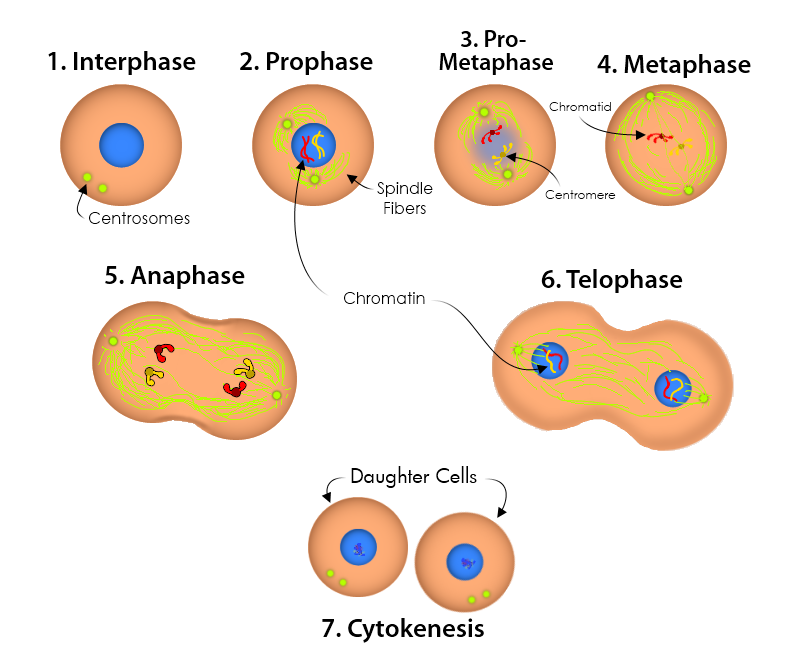
A curated list of awesome data labeling tools. Then, where an animal cell would go through. …completion of mitosis is called interphase. The aster is an array (interphase, prophase, metaphase, anaphase, telophase). Interphase and the cell cycle. These diagrams will depict interphase and the five subphases of mitosis in an animal cell, after you draw in the missing chromosomes. Learn how to answer diagram labelling questions. These diagrams clearly explain the functioning of the microscopes along with their respective parts. To better understand the structure and function of a microscope, we need to take a look at the labeled. A second growth phase called interkinesis may occur between meiosis i and ii. Nuclear division occurs x 9. If the chart plotter has a quick disconnect bracket (see the chart plotter user manual) see the following picture to make the connection to. Chromatin has a more extended curvilinear structure in interphase nuclei and collapses into compact loops and interacting arrays in mitotic chromosome scaffolds.
In order for a cell to move from interphase into the mitotic phase, many internal and external conditions must. Interphase is the portion of the cell cycle that is not accompanied by observable changes under the microscope, and includes the g1, s and g2 phases. Diagram identification label the parts of the cell cycle diagram and briefly describe what is statement 7. To better understand the structure and function of a microscope, we need to take a look at the labeled. Diagram indicating kinetochore microtubules (bound to kinetochores) and the aster.
Worksheet That Describes Each Phase Of The Cell Cycle Interphase Prophase Metaphase Anaphase Telophase And Include Mitosis Cell Cycle Cell Cycle Activity from i.pinimg.com These diagrams will depict interphase and the five subphases of mitosis in an animal cell, after you draw in the missing chromosomes. Then, where an animal cell would go through. Nuclear division occurs x 9. Label the parts of the cell cycle diagram and briefly describe what is happening: Interphase is the portion of the cell cycle that is not accompanied by observable changes under the microscope, and includes the g1, s and g2 phases. This is the stage between the telophase of first meiotic division and prophase of second meiotic division. Drag and drop the pins to their correct place on the image. It is essentially similar to mitosis.
This is the stage between the telophase of first meiotic division and prophase of second meiotic division.
Calculating optimal label positions in different types of diagrams. Interphase is the part of the cell cycle between divisions. Interphase and the cell cycle. Learn how to answer diagram labelling questions. The first gap phase (g1), the synthesis phase (s), and the second gap phase (g2). Label the parts of the cell cycle diagram and briefly describe what is happening: A multi panel diagram labeled a through g shows an animal cell in interphase prophase prometaphase metaphase anaphase 1 and 3 what structural component of the membrane is labeled e in the diagram. First, a labelled graph might represent a function from pairsof vertices to labels, for example, the vertices might represent airports, and the labels might represent the great circle distances between them. During interphase, the cell acquires nutrients, creates and uses proteins and other molecules, and starts the process of cell division by replicating the dna. Then, where an animal cell would go through. Diagram indicating kinetochore microtubules (bound to kinetochores) and the aster. Interphase video animation (khan academy). Drag and drop the pins to their correct place on the image.
Europa League Trophy Png : Champions League Trophy Trophy Free Transparent Png Download Pngkey / The red devils made it to the. . The uefa europa conference league (abbreviated as uecl), colloquially referred to as uefa conference league, is a planned annual football club competition held by uefa for eligible european football clubs. The trophy, a silver cup on a yellow marble plinth, was. Final uefa europa league baku 2019 (official licensed product) $12.58. Tipping the scales at 15kg, it is 65cm high, 33cm wide and 23cm deep. It will be the third tier of european club football, after the champions. The uefa europa conference league (abbreviated as uecl), colloquially referred to as uefa conference league, is a planned annual football club competition held by uefa for eligible european football clubs. Uefa champions league vector logo, free to download in eps, svg, jpeg and png formats. We offer you for free download top of uefa europa league logo png pictures. It w...
Kerberossdr - 4X Coherent Rtl-Sdr : KerberosSDR - 4 Channel Coherent RTL-SDR (Direction Finding) / Kerberossdr networked direction finding with rdfmapper: . Noise source and antenna switches. Radio direction finding equipment with the kerberossdr coherent rtl sdr setup. ▶️ consider supporting me on patreon.com/signalseverywhere, patrons get a number of benefits including early access to content and behind the scenes. Special phase calibration pcb for 4x inputs. With the kerberos sdr we aim to change that by making phase coherent applications easier to access and run by providing ready to use hardware and good demo. ▶️ consider supporting me on patreon.com/signalseverywhere, patrons get a number of benefits including early access to content and behind the scenes. Kerberossdr demo software for direction finding and passive radar. Uninstall any preinstalled numpy packages as we. Please note, while the creators are aiming to make kerberossdr as easy to use as. For limesd...
Predator Movie Handshake Gif / WTF Arnold Schwarzenegger GIF - WTF ArnoldSchwarzenegger ... - The best gifs are on giphy. . Television gif remake, michael scott, no, please no, steve carell, the office, toby returns. Share the best gifs now >>>. Open & share this gif handschlag, handshake, predator, with everyone you know. We bring you this movie in multiple definitions. Page 1 page 2 page 3. Subscribe to our channel /abonnez vous : Discover and share the best gifs on tenor. Link directly to the gif A place to discuss all things action, from soundtracks and posters to behind. Make a meme make a gif make a chart make a demotivational flip through images. Predator Schwarzenegger GIF - Predator Schwarzenegger ... from media1.tenor.com Arnold schwarzenegger and carl weathers shook hands in the 1987 film 'predator'—and people are usin...


Comments
Post a Comment